"Dental Implants Net" is an information site aimed at providing a better public understanding and popularization of dental implants.
Why does the bone quantity become deficient?
Implant treatment requires sufficient bone width and depth at the site for placement. However, there are many cases where the results of dental examination reveal the inability to undergo implant treatment due to the insufficient bone quantity. What does it mean for the bone quantity to "become deficient"? Why does the bone quantity become deficient?
The major causes of bone deficiency are:
- 1. gum disease (alveolar pyorrhea),
- 2. congenital bone deficiency,
- 3. prolonged period of tooth loss, and
- 4. ill-fitting dentures.
- 1. Gum disease (alveolar pyorrhea)
Gum disease, i. e. alveolar pyorrhea, causes resorption of the alveolar bone supporting the tooth, resulting in tooth loss.
Bone resorption proceeding to tooth loss indicates considerable alveolar bone loss.
- 2. Congenital bone deficiency
Congenital bone deficiency includes cases with the thin upper jawbone. In such cases, if an implant is placed, the tip of the implant may perforate the maxillary sinus.
- 3. A prolonged period of tooth loss
If the period of tooth loss is prolonged, adequate stimulation cannot be transmitted to the bone. The human body shows a decrease in function without stimulus. Loss of the jawbone represents bone resorption. If no adequate stimulation is give to the bone, new bone cannot be generated to make up for bone resorption, resulting in bone deficiency.
- 4. Ill-fitting dentures
ll-fitting dentures also cause bone loss. If the adaptation of the dentures is poor, pressure is applied to the gum from various directions and the jawbone (alveolar bone) under the dentures is resorbed by compression.
How can bone deficiency be resolved?
Can implant treatment be performed even with bone deficiency? Can anything be done about congenital bone deficiency? Treatments are available to make up for bone deficiency.
What can the treatments available to make up for bone deficiency?
In cases of bone deficiency, treatment to increase the bone is performed before implant placement. For detail, refer to the linked page with illustrations.
GBR (Guided Bone Regeneration)

The GBR technique is used to facilitate bone regeneration when the bone thickness in the jaw is insufficient for implant placement.
After implant placement, the area of bone deficiency is packed with autogenous bone (i.e., the patient’s own bone) and/or an artificial bone replacement material and covered with a special membrane. Then, osteoblasts are produce to generate new bone within 3 to 4 months. If a small amount of autogenous bone is needed, the bone is collected from the mentum (the lower anterior portion). If a large amount of autogenous bone is required, it is collected from the iliac bone (the hipbone).
Sinus lift

The sinus lift technique is used in cases in which the height and width of the upper jaw are extremely insufficient. The sidewall of the upper gum is cut to expose the bone surface and form a window of about 10 – 30 mm2 into the alveolar bone. Then, the alveolar bone and Schneiderian membrane (the mucosa between the alveolar bone and maxillary sinus) are separated carefully to pack the grafting bone. Over the following 3 – 6 months, the bone is formed. Thereafter, as an implant is placed. The treatment takes about 9 months.
Socket lift

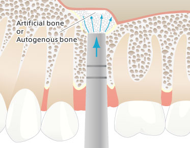
The socket lift technique is a method for elevation of the maxillary sinus with a special tool called an osteotome due to the insufficient bone height and is applicable in cases where the bone height to the Schneiderian membrane is greater than 5 mm. The Schneiderian membrane is pushed up gradually to obtain sufficient bone thickness while packing grafting bone into the socket (the site where the tooth has developed) made for implant placement. The implant is placed as soon as socket lift is completed. The treatment takes about 4 months before it is possible to chew normally.
Unlike the socket lift technique, the sinus lift technique does not increases the bone height by packing grafting materials from the lateral side of the maxillary sinus, but pushes up the maxillary sinus from the sinus floor.
Even difficult cases can be resolved. It is important to have counseling sessions with dentists before deciding on implant treatment.

- What type of dental treatment do you require?
- Basic knowledge of dental implants
- Dental Implants surgery
- Before&After - Photos of dental implant cases
- Success stories
- FAQ
- Privacypolicy
- About us
- sitemap
- Dental Glossary
- Find an Implant Dentist
- To dentists and managers


